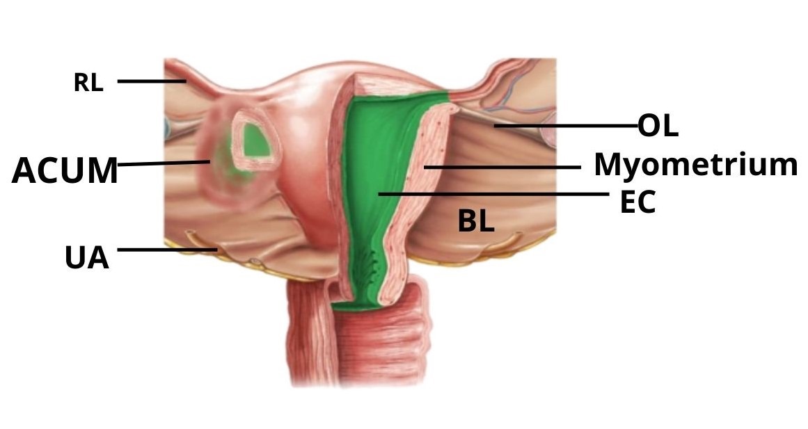
Accessory Cavitated Uterine Mass
Overview
An Accessory Cavitated Uterine Mass (ACUM) is a rare developmental (congenital) uterine anomaly.
It occurs when a small, isolated pocket of uterine tissue — lined with the same type of cells as the uterus — forms outside the main uterine cavity, usually near the round ligament.
Although this tissue looks normal, it does not communicate with the main uterine cavity. During menstruation, this cavity also bleeds internally, leading to pain and swelling.
ACUM is a developmental anomaly which is not cancerous.
Who Can Be Affected?
• Usually diagnosed in young women (teens or early 20s)
• Often have normal menstrual cycles
• Pain usually starts soon after menarche (first period)
Symptoms
● Severe Dysmenorrhoea
● Pelvic pain
● Pain may not respond well to painkillers
● Occasionally, a palpable mass or feeling of fullness on one side
Diagnosis
Ultrasound (USG)
Shows a well-defined cystic mass near the uterus, often on one side.
MRI (Magnetic Resonance Imaging)
Best imaging method to confirm diagnosis.
MRI shows a small cavity within a mass containing blood (similar to menstrual blood), separate from the main uterine cavity.
Laparoscopy
Both diagnostic and therapeutic.
Confirms the presence of a separate cavity and allows for surgical removal.
Management
- Medical Treatment
Usually ineffective, as the trapped blood cannot drain.
Pain may be temporarily controlled with hormonal medicines (oral contraceptives, GnRH analogues), but symptoms often return. - Surgical Treatment
Laparoscopic excision is the treatment of choice.
The accessory mass is carefully removed, and the normal uterus is preserved.
Surgery gives permanent relief from pain and allows normal future fertility.
Postoperative Care
Usually a short hospital stay (1–2 days).
Pain relief medications and routine wound care.
Follow-up after 1–2 weeks, and ultrasound if needed.